Longevity
Blood proteins may flag Alzheimer's risk earlier than spinal fluid, monkey study finds
By Life and Health Today Staff, . Life and Health Today.
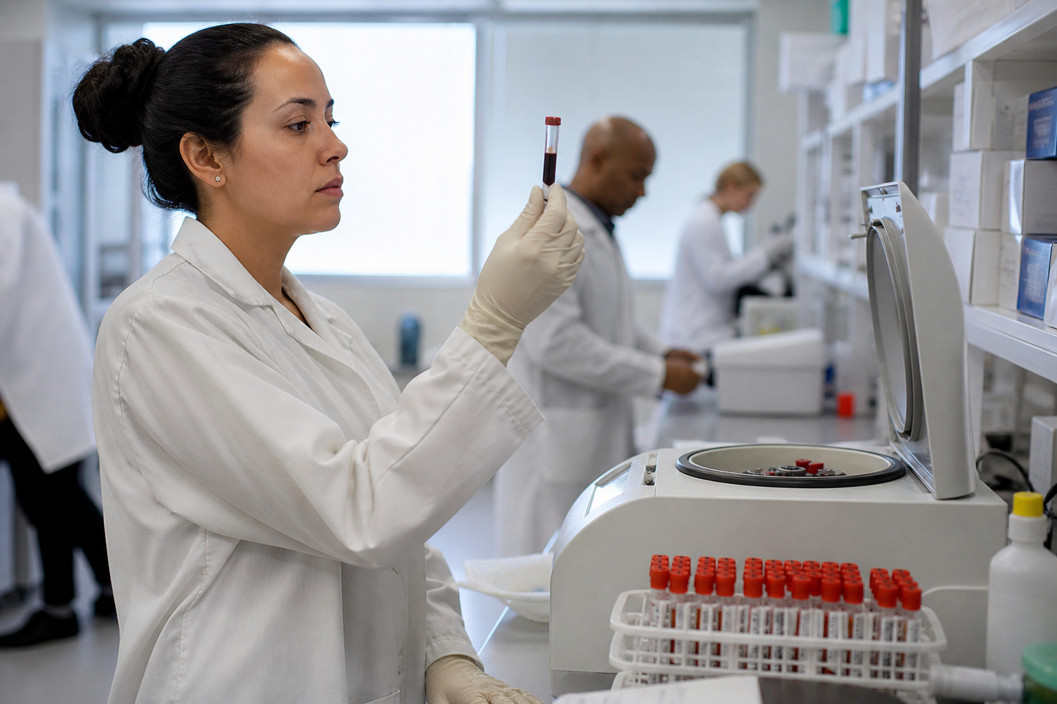
A study published in GeroScience has found that proteins circulating in the blood of aging rhesus macaques may reflect the buildup of amyloid in the brain more faithfully than the same proteins measured in cerebrospinal fluid, the liquid that bathes the brain and spinal cord.
The researchers used a proteomic platform called NULISAseq, which measures many proteins simultaneously, to profile blood plasma and cerebrospinal fluid from 66 rhesus macaques spanning three life stages: adult, pre-geriatric, and geriatric. They then compared those results against human reference samples. All of this work was done in non-human primates; no human patients were enrolled in the study.
Amyloid-beta and phosphorylated tau, two proteins whose abnormal accumulation in the brain is a hallmark of Alzheimer's disease, were higher in the blood of older animals and tracked more closely with the degree of amyloid deposited in brain tissue, confirmed by direct examination of the tissue under a microscope, than the same proteins measured in cerebrospinal fluid. That finding matters because drawing blood is far less invasive than a lumbar puncture, the procedure used to collect cerebrospinal fluid.
The study also found that several proteins already used as markers of brain aging in humans behaved similarly in the macaques. Neurofilaments, GFAP, and TREM2, proteins associated with damage to nerve fibers, support cells called astrocytes, and immune cells in the brain called microglia respectively, all rose with age in patterns the authors described as resembling what is reported in human aging. Inflammatory proteins including interleukin-6 and GDF15 were also higher in older animals, a pattern the authors said resembled human inflammaging, the low-grade chronic inflammation associated with getting older.
Not everything moved in the same direction. Proteins linked to synaptic function, the chemical signaling between nerve cells, were lower in the cerebrospinal fluid of older animals, while certain proteins associated with the choroid plexus, a structure in the brain that produces cerebrospinal fluid, were lower in pre-geriatric animals. The authors noted that age-related protein changes were broader and more numerous in blood plasma than in cerebrospinal fluid.
One of the more striking findings was that a subset of geriatric animals clustered together based on their cerebrospinal fluid protein profiles in a way that chronological age alone did not explain. That suggests biological aging in the brain does not proceed at a uniform rate even among animals of similar age, a pattern familiar from human studies but not previously mapped this way in non-human primates.
The GeroScience article was also indexed by PubMed. The full text of the GeroScience publication was not available in its entirety in the sources used for this report; limitations stated by the authors in the latter portion of the paper are not reflected here.
What this study does not establish is whether any of these protein signatures can predict who will develop dementia, or whether intervening to change them would alter the course of disease. The macaque model is useful precisely because the animals develop amyloid deposits naturally without the widespread tau-driven neurodegeneration seen in late-stage Alzheimer's, which allows researchers to study early pathology in isolation. But a biomarker that tracks a disease process is not the same as a biomarker that predicts clinical outcomes in people, and no human trial has yet tested whether these specific plasma signals translate.
The open question is whether the protein patterns identified here will hold up in large human cohorts and, if they do, whether a blood test built around them could identify people at risk early enough for any future intervention to matter. That would require prospective human studies, which this work does not provide.